SG99 HIGH PREMIUM COLOR DOPPLER

Feature​ :

23.8” HD LED Monitor

Sono VF


Auto Calc

SWE


Virtual Face

Panoramic Imaging


3D/4D Smart Face

MI/TI ≤1


Easy Photo Transfer

Ultra Remote

15.6″ Adjustable Touchscreen

Intelligent Doppler


Sono Crystal

HD Zoom

Sono OB (Optional)


4 Probe Connector

Transvaginal 4D (Optional)

TEE Probe Adult & Pediatric (Optional)

Kategori:

Spec :

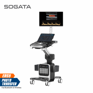
Rotatable : -90°~90°, Foldable : 0°~90°

Rotatable Keyboard: -45°~45°

Flexible Height Adjusment

4D Package + Virtual HD


500GB SSD, DVD-R/W

Imaging Modes: B , 2B , 4B , B/M , B/BC,

CFM,PW, Power Doppler / Directional PD

Instant Triplex, Duplex, Quadplex,


Trapezoidal, Intelligent Doppler

Super Image Module: FHI, Multiple

Compound Imaging, SRA ( Speckle

Reduction Algorithm ), AIO


Q-Image, Q-flow, Q-beam, X contrast

(Intelligent Image Optimization)


Measurement & Calculation Software Packages:

General, OBGYN, Cardiac


Color Doppler Package:

Elastography, Super Needle, 2D Steer, IMT, DICOM 3.0


Cardiac Package :

ECG License, CW, Free Steering M Mode, Color M Mode, TDI, Stress Echo

Images Quality :


Probes :

Volume
Volume
V2-6 (2.5MHz-6.8MHz)
(Optional)
Convex
Convex
C1-5 (2.0MHz-6.8MHz)
Linear
Linear
V4-10 (4.0MHz-12.0MHz)
Transvaginal
Transvaginal
D2D16L (2.0MHz)
(Optional)
Pencils
Pencils
L4-10 (4.0MHz-15.0MHz)
(Optional)
Phassed Array Adult
Phassed Array Adult
S1-5P (1.5MHz-5.3MHz)
(Optional)
Transectal
Transectal
S2-8 (2.0MHz-9.6MHz)
(Optional)


MITI ≤1
MITI ≤1
World standards of safety ultrasound examination recommend to do Mechanical Index (MI) and Thermal Index (TI)
must be ≤1.

The energy released by ultrasound equipment will cause cavitation
effects or the formation of micro
air bubbles that can damage cells,
especially during the embryological
period.

SWE
SWE
SWE is applied to diffuse lesions, providing pressure values to determine lesions
such as fatty liver, liver fibrosis, and
cirrhosis The detection depth can reach
6 centimeters
RT IMT
RT IMT
Simultaneously measure the thickness of the anterior and posterior walls of blood vessels when scanning.
Sono PW
Sono PW
The sample gate can be extended to 4
during PW mode. It can switch and
active each sample gate to realize
multi-point spectrum measurement
in the same cardiac cycle at the same scanning plane
Volume Flow
Volume Flow
A three-dimensional technology which can provide the real-time visualization of hemodynamics blood flow with
a density appearance.