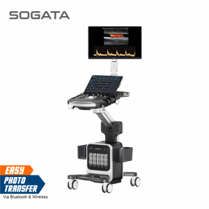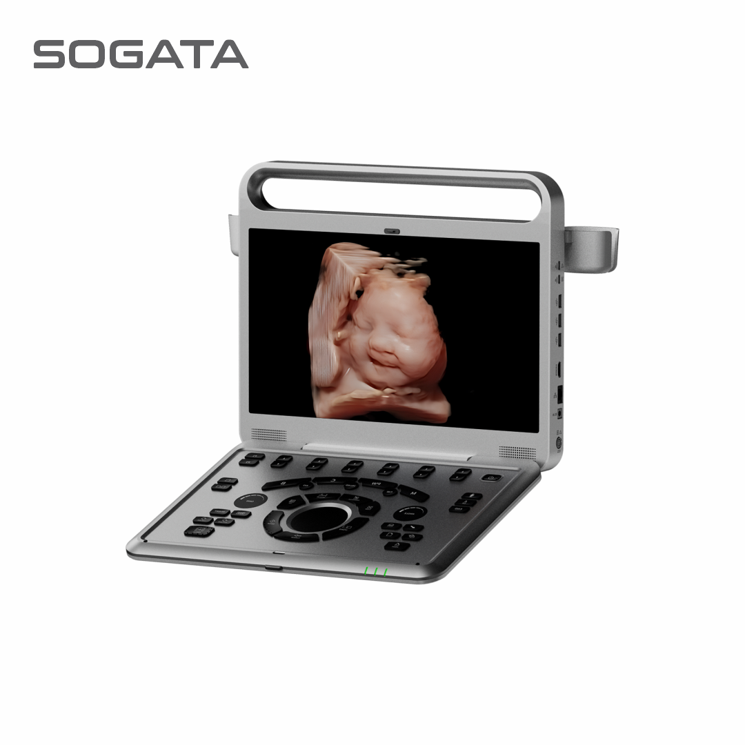
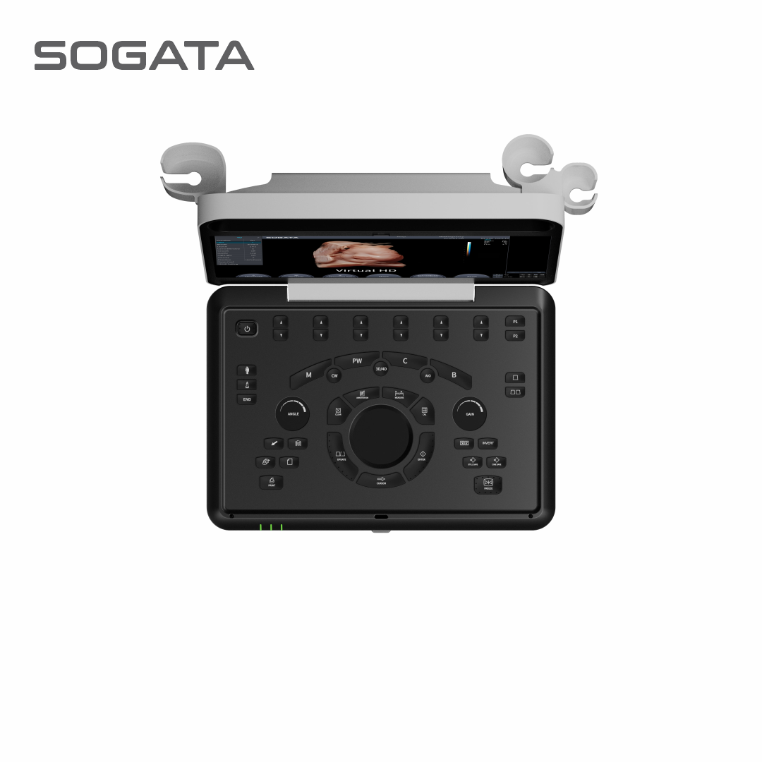
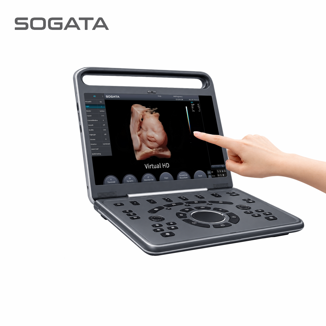
SG90S CD 4D Live HD

Feature


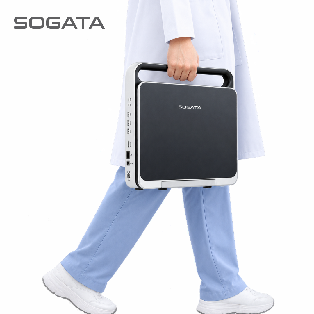
Slim and Lightweight ≤5kg
15,6” Full Touch Display
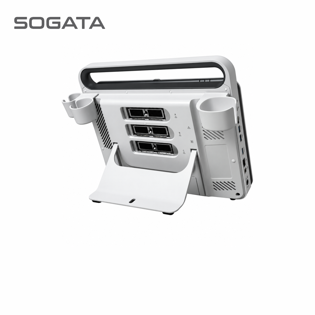
3 Probe Connector Active


Trackpad Keyboard
Sono OB
Virtual HD


Internal Battery
Bluetooth & Wireless
2 Type C Port, HDMI


Automatic PW Trace
Chroma B, M, PW
MI/TI ≤1

Kategori:

Spec

3 Transducer Connectors

3 USB Port & 2 Type C

4D Package + Virtual HD


512GB Hard Disk, HDMI

Imaging Modes: B , 2B , 4B ,

B/M , B/BC, CFM,PW,


Power Doppler / Directional PD

Instant Triplex, Duplex, Quadplex,

Trapezoidal, Full Screen Mode

Automatic PW Trace


Super Image Module:

FHI, Multiple Compound Imaging,

SRA ( Speckle Reduction Algorithm ), Q-Image, Q-flow,

Q-beam, X contrast (Intelligent Image Optimization)


Measurement & Calculation Software Packages:

General, OBGYN, Cardiac


Cardiac Package (Optional) :

ECG License, CW, Free Steering M Mode Color M Mode,

TDI, Stress Echo


Color Doppler Package:

Elastography, Curved Panoramic Imaging, Super Needle 2D Steer,

IMT, DICOM 3.0


Image Quality SG90S CD 4D Live HD

Probe Option SG90S CD 4D Live HD

Volume
Volume
V2-6-D (2.0MHz-5.3MHz)
Convex
Convex
C2-6-D (2.0MHz-5.3MHz)
Transvaginal
Transvaginal
E4-10-D (4.0MHz-10.0MHz)
(Optional)


MITI ≤1
MITI ≤1
World standards of safety ultrasound examination recommend to do Mechanical Index (MI) and Thermal Index (TI)
must be ≤1.

The energy released by ultrasound equipment will cause cavitation
effects or the formation of micro
air bubbles that can damage cells,
especially during the embryological
period.

Elastography
Elastography
Elastography displays tissue stiffness in real time to provide
doctors with addtional diagnostic information when scanning
organs like liver and breast
Virtual HD
Virtual HD
The latest innovation in real-time 4D with powerful imaging engine Greatly streng
then the bond between mother and fetus with moveable virtual light source
Sono OB
Sono OB
Automatically recongnize fetal face. Automatically remove the tissues covering the face. Fast to get fetal face.
Sono Crystal
Sono Crystal
Provide context and surface information. Allow an accurate understanding of patient anatomy. Easily differentiates between soft tissue and structure.
MVI
MVI
It applied a brand- new algorithm to check the blood flow. Greatly improve the visualization sensitivity of low velocity and small blood vessels. Provide more and accurate blood flow information for clinic.