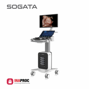
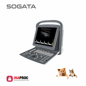
SG60P Cardiac Package 2 Probes

Feature :

Adjustable LED Monitor (0~30°)
Ultra Compact
Easy Photo Transfer (Via Wireless)


One Key Quick Save
Curved Panoramic
Continous Wave (CW) Doppler


15” HD LED Monitor
4 USB Port, VGA, S-Video
Automatic PW Trace


Free Stering M-Mode
Chroma B, M, PW
MI/TI ≤1


Category:

Spec


2 Transducer Connectors
500GB Hard Disk, 4 USB Port
Imaging Modes: B , 2B , 4B ,


B/M , B/BC, CFM,PW,
Power Doppler / Directional PD
Instant Triplex, Duplex, Quadplex,


Trapezoidal, Full Screen Mode
Automatic PW Trace
Printer BW, Printer Computer, UPS, Stabilizer, Trolley


Super Image Module: FHI, Multiple
Compound Imaging, SRA ( Speckle
Reduction Algorithm ),


Q-Image, Q-flow, Q-beam, X contrast
(Intelligent Image Optimization)


Measurement & Calculation Software Packages:
General, OBGYN, Cardiac


Cardiac Package:
ECG License, CW, Free Steering M Mode
Color M Mode, TDI, Stress Echo


Color Doppler Package:
Elastography, Curved Panoramic
Imaging, Super Needle 2D Steer,
IMT, DICOM 3.0


Image Quality SG60P CDCP

Probe SG60P CDCP

Phassed Array Adult
Phassed Array Adult
S1-5-D (1.0MHz-5.3MHz)
Linear
Linear
L3-E-10D (4.0MHz-10.0MHz)
Convex
Convex
C2-6-D (2.0MHz-5.3MHz)
(Optional)
Phassed Array Pediatric
S2-8-D (2.0MHz-8.0MHz)
(Optional)


MITI ≤1
MITI ≤1
World standards of safety ultrasound examination recommend to do Mechanical Index (MI) and Thermal Index (TI)
must be ≤1.

The energy released by ultrasound equipment will cause cavitation
effects or the formation of micro
air bubbles that can damage cells,
especially during the embryological
period.

Tissue Doppler Imaging (TDI)
Tissue Doppler imaging is a novel echocardiography technique that
directly measures myocardial velocity.

Systolic TD measurements assess left and right ventricular myocardial contractile function. Diastolic TD values reflect myocardial relaxation.
Free steering M mode
Tissue Doppler imaging is a novel echocardiography technique that
directly measures myocardial velocity.

Systolic TD measurements assess left and right ventricular myocardial contractile function. Diastolic TD values reflect myocardial relaxation.
Sono IMT
Sono IMT
Auto trace/Manual trace. Monitor the IMT regulary,
analysis the data which is helpful for chronic disease management.
Sono PW
Sono PW
The sample gate can be extended to 4 during PW mode.

It can switch and active each sample gate to realize
multi-point spectrum measurement in the same cardiac
cycle at the same scanning plane; Support synchronous
display of spectrum and speed value.
Stress Echo
Stress Echo
Help to confirm or rule out the presence of coronary artery disease.

Patients with coronary artery blockages may have
minimal or no symptoms during rest.

Symptoms and sign of heart disease may be unmasked
by exposing the herart to the stress of exercise.