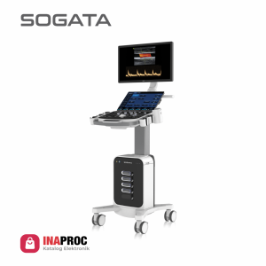
SG8 Cardiac Package 3 Probes

21.5” HD LED Monitor

Phassed Array Adults & Pediatric
6 USB Port, VGA, S-Video


TDI, CW
Chroma B, M, PW
Automatic PW Trace

Stress Echo
Free Stering M-Mode
MITI ≤1


Curved Panoramic
MI/TI ≤1


Easy Photo Transfer
Curved Panoramic
Ergonomic


Continuous Wave (CW) Doppler
Auto IMT
Hero KIT


4 Probe Connector Active
Smart Standby Mode

Color M Mode
TEE Probe T5P64L Adult & Pediatric (Optional)

Link SG8 CDCP 3 Probes :

Category:

Spec :


Rotatable : -90°~90°,
Foldable : 0°~90°


500G Hard Disk, DVD-R/W


Imaging Modes: B , 2B , 4B , B/M,
B/BC, CFM,PW, Power Doppler /
Directional PD


Instant Triplex, Duplex, Quadplex,
Trapezoidal,

Sony BW, Printer Color, Stabilizer, UPS


Super Image Module: FHI, Multiple
Compound Imaging, SRA ( Speckle
Reduction Algorithm ), AIO


Q-Image, Q-flow, Q-beam, X contrast
(Intelligent Image Optimization)


Measurement & Calculation Software Packages:
General, OBGYN, Cardiac


Color Doppler Package:
Elastography, Curved Panoramic, Super Needle 2D Steer, IMT, DICOM 3.0


Image Quality SG8 Cardiac Package

Probe SG8 Cardiac Package

Convex
Convex
D3C60L (2.0MHz-6.8MHz)
Linear
Linear
D12L40L (7.0MHz-18.0MHz)
Transvaginal
Transvaginal
D7C10L (4.0MHz-15.0MHz)
(Optional)


MITI ≤1
MITI ≤1
World standards of safety ultrasound examination recommend to do Mechanical Index (MI) and Thermal Index (TI)
must be ≤1.

The energy released by ultrasound equipment will cause cavitation
effects or the formation of micro
air bubbles that can damage cells,
especially during the embryological
period.

Stress Echo
Stress Echo
Help to confirm or rule out the presence of coronary artery disease.

Patients with coronary artery blockages
may have minimal or no symptoms during rest.

Symptoms and sign of heart disease
may be unmasked by exposing the herart
to the stress of exercise.
Quantative Elastography
Display the elasticity of different tissues in different color. Provide more clinical information, especially for soft tissue.

Strain ratio measurement quantitatively
gives the ratio between the average strain
of the selected region and of the nearby normal tissue region.
Tissue Doppler Imaging (TDI)
Tissue Doppler imaging is a novel echocardiography technique that
directly measures myocardial velocity.

Systolic TD measurements assess left and right ventricular myocardial contractile function. Diastolic TD values reflect myocardial relaxation.
Free steering M mode
Obtain accurate cardiac function analysis data. Obtain accurate cardiac measurement parameters of any section and any Angle.

Excellent and convenient for difficult patient examination.