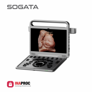
SG60P Color Doppler 2 Probes

Feature

Adjustable LED Monitor (0~30°)
Ultra Compact
Easy Photo Transfer (Via Wireless)


One Key Quick Save
Curved Panoramic
Virtual HD (Optional)


15” HD LED Monitor
Shortcut Key Function
4 USB Port, VGA, S-Video


Automatic PW Trace
Chroma B, M, PW
MI/TI ≤1

Category:

Spec
2 Transducer Connectors
500GB Hard Disk, 4 USB Port

4D Package (Optional)
Virtual HD (Optional)


Imaging Modes: B , 2B , 4B ,
B/M , B/BC, CFM,PW,
Power Doppler / Directional PD


Instant Triplex, Duplex, Quadplex,
Trapezoidal, Full Screen Mode
Automatic PW Trace
Printer BW, Printer Computer, UPS, Stabilizer, Trolley


Super Image Module: FHI, Multiple Compound Imaging,
SRA ( Speckle Reduction Algorithm ), Q-Image, Q-flow,
Q-beam, X contrast (Intelligent Image Optimization)


Measurement & Calculation Software Packages:
General, OBGYN, Cardiac


Cardiac Package (Optional) :
ECG License, CW, Free Steering M Mode
Color M Mode, TDI, Stress Echo


Color Doppler Package:
Elastography, Curved Panoramic
Imaging, Super Needle 2D Steer,
IMT, DICOM 3.0


Image Quality SG60P Color Doppler

Probe SG60P Color Doppler
Linear
Linear
L3-10-D (4.0MHz-10.0MHz)
Convex
Convex
C2-6-D (2.0MHz-5.3MHz)
Transvaginal
Transvaginal
E4-10-D (4.0MHz-10.0MHz)
(Optional)


MITI ≤1
MITI ≤1
World standards of safety ultrasound examination recommend to do Mechanical Index (MI) and Thermal Index (TI)
must be ≤1.

The energy released by ultrasound equipment will cause cavitation
effects or the formation of micro
air bubbles that can damage cells,
especially during the embryological
period.

Elastography
Elastography
Elastography displays tissue stiffness in real time to provide
doctors with addtional diagnostic information when scanning
organs like liver and breast
Virtual HD (Optional)
The latest innovation in real-time 4D with powerful imaging engine Greatly streng
then the bond between mother and fetus with moveable virtual light source